Tissue Mechanics: Bone and Ligament Biomechanics
Tissue biomechanics class BME 615
Outline
Bone composition and structure
Bone properties: elasticity, strength, and toughness
Bone properties: effect of age and species
Bone properties: anisotropy
Bone properties: viscoelasticity
Bone nonlinearity compared with other tissues
Bone as a natural composite
Bone remodeling and adaptation
Bone structure and properties
The left image shows structure of a cross section of bovine plexiform bone; the center image, of a cross section of human Haversian bone under polarized light.
The far right image shows a stained section of bone, showing the osteocytes (bone cells). The image, after Professor Peter Muir of Veterinary Medicine, is a composite image, obtained with a confocal microscope in bright light, of a decalcified transverse section of the mid-diaphysis of a dog humerus, stained with silver to highlight the canaliculi.
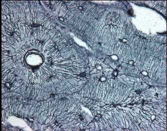
The small dark circles in the human bone at center are cross sections of Haversian canals. The crossed polarizers give rise to the dark color in the empty canals. The large circles are cross sections of osteons, which are large fibers about 200 microns across. Bovine plexiform bone has a laminated structure. Colors in the polarized light images arise from the fibrous structure of bone; the anisotropy gives rise to a wavelength dependent rotation of the polarization of the light.
The images below show schematic diagrams of hierarchical structure of bone and of tendon.
larger image
Hierarchical structure of compact bone. Reference: Lakes, R. S., "Materials with structural hierarchy", Nature, 361, 511-515 (1993).
link
Hierarchical structure of tendon.
Reference: J. Kastelic, A. Galeski, and E. Baer, The Multicomposite Structure of Tendon, Connective Tissue Research, 1978, Vol 6, pp. 11-23.