Mechanical properties of cancellous bone
A review.
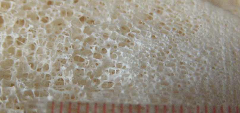
Cancellous or trabecular bone is a highly porous or cellular form of bone. The image shows a photograph of cancellous bone from the leg bone of an animal. The scale marks are in millimeters. In a typical long bone, the cortex or exterior of the shaft (diaphysis) and flared ends (metaphysis) is composed of compact bone while the interior, particularly near the articulating ends, is filled with cancellous bone. Cancellous bone may also be found to fill the interior of short bones and flat bones as well as in the interior of bony tuberosities under muscle attachments. The structure of cancellous bone is that of a latticework of bars and plates. The volume fraction of solid material can be from 5% to 70%; the interstices in living bone are filled with marrow. The compressive strength
s
ult[in
MPa] depends very much on the density
r
(in g/cm
3)
and also varies with the strain rate d
e/dt
(in s
-1)
as follows (3.1).
sult
= 68 [d
e/dt]0.06
r2
This relation also models the compressive strength of compact bone
(221 MPa, at a
density of
r = 1.8 g/cm
3).
Diagram: bone
Modulus, strength vs. density.
To a certain degree of approximation, both compact and cancellous bone may be mechanically viewed as a single material of variable density (3.1). Density is not, however, the only determinant of the properties of cancellous bone. The microstructure can vary considerably from one part of the body to another (3.2). For example, in the vertebrae and in the tibia (3.3), a highly oriented, columnar architecture is observed. This kind of trabecular bone is highly anisotropic: the Young's modulus in the longitudinal direction can exceed that in the transverse direction by more than a factor of ten (3.3). By contrast, in regions such as the proximal part of the bovine humerus, the cancellous bone can be essentially isotropic (3.4). This bone is about twice as strong in compression as in tension. In many ways cancellous bone (3.5, 3.6, 3.7) is similar in its behavior to man-made rigid cellular foams (3.8). For example, in compression, the stress-strain curves contain a linear elastic region, up to a strain of about 0.05, at which the cell walls bend or compress (3.8). A plateau region of almost constant macroscopic stress is associated with elastic buckling, plastic yield, or fracture of the cell walls. The compressive failure of the cancellous bone proceeds at approximately constant stress until the cell walls touch each other; at this point any further compression causes the stress to rise rapidly (3.8). By contrast, fracture of cancellous bone in tension proceeds abruptly and catastrophically (3.4). The
energy absorption capacity of cancellous bone is consequently much less in tension than it is in compression (3.5). This suggests that tensile and
avulsion fractures of cancellous bone observed clinically are associated with minimal energy absorption and therefore may be precipitated by relatively minor
trauma (3.5). The elastic modulus E for cancellous bone increases as the square of the
density
(E = k r2)
if the structure consists of open cells forming a network of rods (3.6, 3.7).
In closed cell cancellous structures consisting of plates, the modulus E is proportional to the cube of the density
(E = k r3).
Based on study of micrographs and density maps of femora and vertebrae, it is suggested that an open cell structure of rods is found when the solid volume fraction is less than about 0.13, while a closed cell plate structure occurs at a density of more than 350 kg/m
3
corresponding to a relative density or solid volume fraction of 0.20 (3.6).
The relationship between density and structure may not be so straightforward in all situations. As for the highly oriented columnar cancellous bone from the human tibia (3.3), the modulus E in the longitudinal direction is proportional to the density
(E= kr).
Such behavior is anticipated on the basis of an axial compressional mode of deformation of the cell walls, in contrast to the bending mode which is
expected in rod and plate structures (3.8).
References
3.1. Carter, D.R. and Hayes, W.C., Bone compressive strength: the influence of density and strain rate, Science 194, 1174-1176, 1976.
3.2. Dyson, E.D., Jackson, C.K. and Whitehouse, W.J., Scanning electron microscope studies of human trabecular bone, Nature,
225, 957-959, 1970.
3.3. Williams, J.L. and Lewis, J.L., Properties and an anisotropic model of cancellous bone from the proximal tibial epiphysis,
J. Biomechanical Engng. 104, 50-56, 1982.
3.4. Kaplan, S., Hayes, W.C., Stone, J.L., and Beaupre, G.S., Tensile strength of bovine
trabecular bone, J. Biomechanics, 18, 723-727, 1985.
3.5. Carter, D.R., Schwab, G.H., and Spengler, D.M., Tensile fracture of cancellous bone,
Acta Orthop, Scand. 51, 733-741, 1980.
3.6. Gibson, L.J., The mechanical behaviour of cancellous bone,
J. Biomechanics , 18,
317-328, 1985.
3.7. Carter, D.R. and Hayes, W.C., The compressive behaviour of bone as a two-phase
porous structure, J. Bone Jnt. Surg, 59A, 954-962, 1977.
3.8. Gibson, L.J. and Ashby, M.F. The mechanics of three-dimensional cellular
materials, Proc. Royal Soc. London A382, 25-42, 1982.
3.9 Gibson, L. J. and Ashby, M. F., Cellular solids, Cambridge, University Press, 1997.
Top